- 501 Orchard Road, #04-08 Wheelock Place, Singapore 238880
- (65) 6235 4429
- hello@vidaskinclinic.com
- (65) 9830 0597
By Dr Kay Low, Senior Aesthetic Physician, VIDASKIN
Most patients know Ultherapy as an ultrasound skin-lifting treatment. Fewer realise that the same ultrasound is used to look beneath the skin before any energy is delivered.
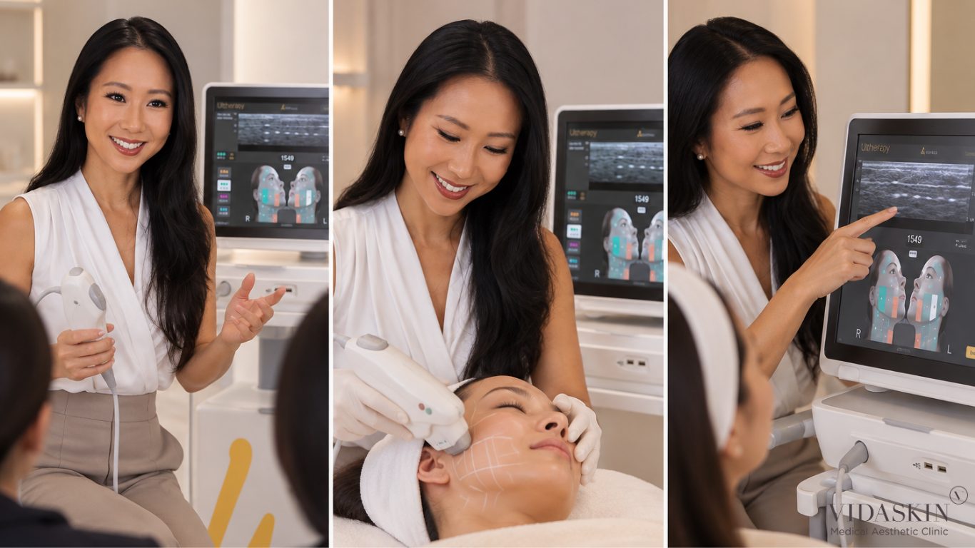
During an Ultherapy treatment, your doctor can see your skin, fat and SMAS on a screen in real time. That image shapes every decision that follows. In this article, I explain what appears on the screen, why your layers are not the same as anyone else’s, and why imaging matters most in the areas patients care about most.
Many energy-based lifting and tightening treatments are delivered without real-time imaging. The practitioner relies on anatomy knowledge, standard depth settings and feel. This can work well in experienced hands, but it means the depth of each person’s tissue is assumed rather than seen.
Ultherapy is different. It combines microfocused ultrasound energy with ultrasound visualisation, so the doctor can confirm what lies beneath the transducer before each line is placed. The newest platform, Ultherapy PRIME, builds on this with an updated imaging display.
On the Ultherapy screen, tissue layers appear as bands of differing brightness:
The SMAS, or superficial musculoaponeurotic system, is the fibrous layer surgeons lift and tighten during a facelift. Ultherapy’s deepest transducer is designed to reach this layer without incisions.
Two people of the same age can have very different layer thicknesses. Skin thickness, fat distribution, muscle tone and bone structure all vary with genetics, weight, hormones, and age.
As an aesthetic doctor with a special interest in women’s skin across life stages, I see this clearly on the scan. Skin and fat distribution can look quite different after pregnancy, with weight change, or through perimenopause. Imaging lets me plan for the face in front of me now, not the average face in a protocol.
The SMAS is not a flat sheet at a uniform depth. Even within one face, it sits closer to the surface in some areas and deeper in others. It is thicker in some zones and thinner, or less distinct, in others. Over the cheekbone, it may sit close to bone. Along the jawline, it may sit deeper beneath fuller fat.
This is why a single depth setting cannot be correct everywhere. The scan lets the doctor match the transducer to the tissue, zone by zone.
If the scan shows the SMAS is shallower than the 4.5 mm setting in a particular zone, or that bone lies close beneath it, the doctor can switch to a shallower transducer or move the treatment line. If the fat is thicker, deep lines may be concentrated where they will engage the SMAS most effectively.
Imaging also shows where energy would not achieve much, such as over bone or where the transducer is not properly coupled to the skin. If the image breaks up, the contact is poor and the line would not deliver energy as intended. Recognising this saves lines for where they matter.
Occasionally, the scan and examination together suggest Ultherapy is not the right choice, or not the right choice yet. Examples include very thin soft tissue over bone throughout, advanced laxity beyond what a non-surgical treatment can address, or volume loss being the main concern.
In these cases, the most useful thing I can do is say so. Sometimes the right recommendation is a different treatment, a combined plan, or simply waiting.
The jawline is where many patients first notice change: softening of the contour and early jowls. Here the SMAS can sit deeper beneath lower-face fat. The marginal mandibular branch of the facial nerve also runs in this region. Imaging helps the doctor engage the right depth along the jaw while respecting the anatomy around it.
Under the chin, fat thickness varies widely between people. In some, the platysma muscle and its overlying tissue are close to the surface. In others, a fuller fat layer sits in between. The scan helps decide how deep lines should be and how much the area can usefully take.
Around the eyes and brow, the skin is thin, and bone lies close beneath. Precise depth selection is essential for comfort, safety, and effectiveness. Imaging helps the doctor stay within safe tissue and outside the orbital rim, using shallower transducers where appropriate.
Many clinics will say their device has imaging. The more useful question is whether imaging is used on you, in your session, before each zone.
You might ask:
A doctor who uses imaging routinely will be comfortable answering these questions and showing you what they see.
Ultherapy treatment is generally best suited to people with mild to moderate laxity who want gradual, natural-looking improvement without surgery. Good candidates often have:
It is less suitable for advanced laxity, significant volume loss, or where surgical-level change is expected.
Ultherapy relies on the body’s own collagen remodelling, so change is gradual. Patients often notice early firmness within weeks, with more visible lifting typically from two to three months. Clinical studies of microfocused ultrasound show measurable improvement in suitable patients, though the effect is more subtle than surgery and varies between individuals.
Imaging does not change the biology of collagen remodelling. What it improves is the precision with which energy reaches the intended tissue, which is a key part of getting a consistent result.
The SMAS (superficial musculoaponeurotic system) is a fibrous layer beneath the skin and fat that connects with the facial muscles. It provides structural support to the face and is the layer surgeons tighten during a facelift.
Yes. Ultherapy’s deepest transducer focuses energy at around 4.5 mm, which corresponds with the SMAS in many areas of the face. Real-time imaging helps confirm the depth in each zone.
It shows the layers beneath your skin: the dermis, subcutaneous fat, the SMAS, and in some areas muscle and bone. This lets your doctor confirm depth and transducer contact before each line.
No. Ultherapy combines treatment with real-time visualisation. Many other focused ultrasound (HIFU) devices treat without imaging, relying on standard depth settings.
Both use focused ultrasound energy. Ultherapy adds real-time imaging, allowing the doctor to see tissue layers and adjust depth before placing each line. Results with any device also depend on the practitioner.
Skin thickness, fat distribution, muscle and bone structure vary with genetics, age, weight and hormones. The SMAS can sit at different depths in different people, and in different zones of the same face.
At VIDASKIN, you are welcome to ask your doctor to show you the scan during your assessment. Seeing your own layers often helps patients understand their treatment plan.
Imaging helps the doctor identify bone, confirm depth, and check transducer contact, which supports more precise and safer energy placement. Safety also depends on anatomical knowledge and avoiding nerve danger zones.
Scanning adds a little time, but it is part of a well-planned treatment. A full face and neck session commonly takes around 60 to 90 minutes, depending on the areas treated.
Most patients feel brief heat and a deep ache as lines are delivered, particularly over bony areas. Comfort varies between people and zones, and your doctor can discuss pain-relief options with you.
No. Ultherapy offers gradual, non-surgical lifting for mild to moderate laxity. It does not replicate the degree of change achieved by surgery, and for advanced laxity, a surgical opinion may be more appropriate.
Often, yes. However, your doctor needs to know where and when fillers were placed so treatment can be planned and sequenced appropriately.
Ultherapy may not be suitable for people with active skin infection or open wounds in the treatment area, certain implants in the area, or during pregnancy. It may also be unsuitable where expectations exceed what non-surgical lifting can achieve. Your doctor will assess this individually.
Some patients notice early firmness within weeks. More visible lifting typically develops from two to three months, with continued improvement for some time after.
Downtime is usually minimal. Mild redness, swelling, tenderness or temporary numbness can occur and generally settle within days to a few weeks. Most patients return to normal activities straight away.
Yes. The neck and submental (under-chin) area are commonly treated to improve laxity and contour. Imaging is particularly useful here because fat thickness varies widely between people.
Ultherapy uses focused ultrasound to reach specific depths, including the SMAS, with real-time imaging. RF skin tightening, such as XERF, uses radiofrequency energy to heat the skin more broadly. The two are sometimes combined because they work at different levels.
Every face has its own layers, and good Ultherapy starts by looking at them. At VIDASKIN, your Ultherapy PRIME treatment is planned by a doctor who scans first, explains what they see, and adjusts the plan to your anatomy.
If you’d like to see your own SMAS on screen, just ask during your assessment.
Book an Ultherapy assessment with Dr Kay Low.
Dr Kay Low is a Senior Aesthetic Physician at VIDASKIN. She graduated in medicine from the University of Cambridge (MA, MB BChir) and is registered to practise in both Singapore and the United Kingdom. Her special interests include early facial ageing in the 30s and 40s, skin quality and women’s skin across life stages.
This article is for general education and does not replace an individual medical consultation. Suitability and results vary between patients.

Dr. Kay Low is a UK-trained medical doctor with over 7 years of clinical experience across the UK and Singapore, specialising in aesthetic medicine. A graduate of the University of Cambridge, she has worked across multiple medical and surgical disciplines in the UK and Singapore before completing postgraduate training in Family Medicine and advanced training in aesthetic medicine.
